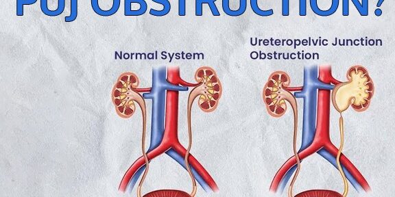
What does PUJ stand for?
PUJ stands for Pelvic-Ureteric Junction. It is a junction where the renal pelvis joins the upper end of the ureter. The ureter is a long thin tubular structure approximately 26cm long through which the urine from the kidney reaches the bladder. The renal pelvis is a funnel shape and has cup-like extensions, called Calyces, within the kidney. Normally the kidney performs its function and removes waste, salt, and excess water from blood in the form of urine and this urine collects within the renal pelvis. Then this urine passes through the ureter and flows into the bladder.
What is PUJ Obstruction?
PUJ Obstruction means the narrowing down of the pelvic-ureteric junction (the site where kidney pelvis joins the upper end of the ureter) due to which the flow of urine from the kidney to the bladder is obstructed. This blockage may cause the flow of urine in a slow manner, or it is completely stopped. This urine due to blockage gets collected in the kidney causing swelling inside the kidney and consequently, damaging the kidney. In case the blockage is significant and there is effect on the kidney due to blockage, surgery may be required to improve the flow of urine and hence preserve the kidney function.
PUJ Obstruction is classified as either:
1) Congenital/Primary – It is present since birth as a defect that could have occurred when the kidneys are in the developmental phase. The cause of developing congenital PUJ Obstruction may be the outcome of the abnormal joining of the ureter into the renal pelvis, abnormal arrangement of ureteric musculature at the PUJ, or a crossing artery running adjacent to the PUJ causing a blockage.
2) Acquired/Secondary – This condition of the PUJ Obstruction may be the result of previous surgery, kidney stones, or trauma.
What are the Causes of PUJ Obstruction?
PUJ Obstruction is seen mostly in infants by birth which means children are born with this condition. One in every 1,500 children are born with this condition. The blockage occurs during the period when the kidneys are forming. With the help of antenatal ultrasound, a doctor often predicts PUJ Obstruction before the child is born.
Symptoms of PUJ Obstruction?
There may not be any symptoms of PUJ Obstruction. When symptoms occur, they are:
1) Abdominal mass.
2) Frequent Urinary Tract Infection.
3) Pain in the upper abdomen or back, mostly felt at the time of fluid intake.
3) Blood in Urine.
4) Kidney Stones
What are the Available Treatments of PUJ Obstruction?
In infants, the treatment is not always necessary, and experts have different opinions. In infants below the age of 18 months, the problem of poor drainage may be temporary. It has been observed that infants with good kidney function, but bad drainage have shown improvement after a few months. On the other hand, in some infants with PUJ Obstruction, this problem will not improve, and situation becomes bad to worst.
Infants with enlarged kidneys are followed by repeat ultrasounds. In the first 18 months of life sudden improvement may occur. In case the urine flow does not improve, and the obstruction is there, then surgery is required. In adults also, evaluation is done with scans, and if PUJ obstruction is definitively diagnosed, then the treatment is carried out.
Open Surgery
The classic treatment is an operation called pyeloplasty. During this surgery, the PUJ is removed, and the ureter is again attached to the renal pelvis aiming to create a wide opening. This way it lets the urine drain easily. Also, it relieves symptoms and the risk of infection. It would take a few hours with a very good success rate of about 95%. After the surgery, the patient may have to stay in the hospital for a day or two.
This operation can be done by open technique when a cut is made in abdomen and surgery is performed. Also, it can be done by laparoscopy or Robotic technique, mentioned as under:
Minimally Invasive Surgery
These are newer surgical options available with less invasion, like:
1. Laparoscopic Pyeloplasty with or without a surgical robot.
2. Internal Incision of the PUJ using a camera and scope inserted through the camera.
Laparoscopic Pyeloplasty: In this procedure, the surgeon works by making a small cut in the abdominal wall. Surgery is performed by inserting small instruments and hence the skin cut is not large.
Robotic surgery involves use of surgical robot which helps perform the laparoscopic surgery with better precision, magnification, and smaller scars. Lesser pain and faster recovery are the advantages. This procedure has shown very successful results. Internal Incision: In this procedure, a wire is inserted through the ureter. The inserted wire is then used to cut the tight and narrow PUJ from the inside. A special ureter drain is left in for a few weeks and then removed. With time the PUJ heals in a better open manner, but the procedure may need to be repeated. Although the success rate of this procedure is less in comparison to open or laparoscopic pyeloplasty, but benefits include less post operative pain, no or smaller external scar and early recovery. However, this option is possible in selective cases of PUJ obstruction.


